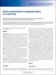
Female sexual dysfunction in androgenetic alopecia: Case-control study
| dc.contributor.author | Sancak, Eyüp Burak | |
| dc.contributor.author | Oğuz, Sevilay | |
| dc.contributor.author | Akbulut, Tuğba | |
| dc.contributor.author | Uludağ, Ayşegül | |
| dc.contributor.author | Akbaş, Alpaslan | |
| dc.contributor.author | Kurt, Ömer | |
| dc.contributor.author | Akbulut, Mehmet Fatih | |
| dc.date.accessioned | 2022-05-11T14:36:59Z | |
| dc.date.available | 2022-05-11T14:36:59Z | |
| dc.date.issued | 2016 | |
| dc.identifier.issn | 1911-6470 | |
| dc.identifier.issn | 1920-1214 | |
| dc.identifier.uri | https://doi.org/10.5489/cuaj.3582 | |
| dc.identifier.uri | https://hdl.handle.net/20.500.11776/8515 | |
| dc.description.abstract | Introduction: We sought to evaluate the association of female sexual dysfunction (FSD) with androgenetic alopecia (AGA) and metabolic syndrome (MetS) in premenopausal women. Methods: From December 2013 to June 2015, we performed a case-control, prospective study of 115 patients with AGA and 97 age-matched control patients without AGA from among premenopausal women who visited dermatology clinics of the two reference hospitals. Comprehensive history, anthropometric measurements, and questionnaire administration were performed for each of the total of 212 women. The Female Sexual Function Index (FSFI) was used to assess the key dimensions of female sexual function. AGA was assessed and graded by an experienced dermatologist according to Ludwig's classification. The MetS assessment was made according to the NCEP-ATP III criteria. Results: In univariate analysis, age, weight, waist circumference, hip circumference, waist-to-hip ratio, body mass index (BMI), AGA, MetS, cardiovascular event, marital status, hypertension, high fasting plasma glucose, high triglyceride, large waist, total testosterone, and free testosterone were associated with presence of FSD. In logistic regression analysis, age (odds ratio [OR] 1.21, 95% confidence interval [CI] 1.13. 1.30; p<0.001), AGA (OR 3.42, 95% CI 1.31. 8.94; p= 0.017), MetS (OR 5.39, 95% CI 1.34. 21.62; p= 0.012), and free testosterone (OR 0.18, 95% CI 0.09. 0.37; p< 0.001) were independently associated with FSD. Conclusions: Our study suggests that age, AGA, MetS, and free testosterone may have strong impact on sexual function in premenopausal women. Further studies with population-based and longitudinal design should be conducted to confirm this finding. | en_US |
| dc.language.iso | eng | en_US |
| dc.publisher | Canadian Urological Association | en_US |
| dc.identifier.doi | 10.5489/cuaj.3582 | |
| dc.rights | info:eu-repo/semantics/openAccess | en_US |
| dc.subject | Middle-Aged Women | en_US |
| dc.subject | Metabolic Syndrome | en_US |
| dc.subject | Insulin-Resistance | en_US |
| dc.subject | Risk-Factors | en_US |
| dc.subject | Index Fsfi | en_US |
| dc.subject | Testosterone | en_US |
| dc.subject | Prevalence | en_US |
| dc.subject | Association | en_US |
| dc.subject | Therapy | en_US |
| dc.subject | Men | en_US |
| dc.title | Female sexual dysfunction in androgenetic alopecia: Case-control study | en_US |
| dc.type | article | en_US |
| dc.relation.ispartof | Cuaj-Canadian Urological Association Journal | en_US |
| dc.department | Fakülteler, Tıp Fakültesi, Cerrahi Tıp Bilimleri Bölümü, Üroloji Ana Bilim Dalı | en_US |
| dc.authorid | 0000-0002-5007-8143 | |
| dc.authorid | 0000-0003-1470-5952 | |
| dc.identifier.volume | 10 | en_US |
| dc.identifier.issue | 7-8 | en_US |
| dc.identifier.startpage | E251 | en_US |
| dc.identifier.endpage | E256 | en_US |
| dc.institutionauthor | Kurt, Ömer | |
| dc.relation.publicationcategory | Makale - Uluslararası Hakemli Dergi - Kurum Öğretim Elemanı | en_US |
| dc.authorscopusid | 55753628300 | |
| dc.authorscopusid | 55749960900 | |
| dc.authorscopusid | 57194558922 | |
| dc.authorscopusid | 55647897100 | |
| dc.authorscopusid | 23977731600 | |
| dc.authorscopusid | 57216014831 | |
| dc.authorscopusid | 56841316400 | |
| dc.authorwosid | Akbulut, Tugba Özkök/AAM-5899-2021 | |
| dc.authorwosid | Akbulut, Mehmet Fatih/N-3886-2019 | |
| dc.authorwosid | Akbas, Alpaslan/AAX-5617-2020 | |
| dc.identifier.wos | WOS:000382528100008 | en_US |
| dc.identifier.scopus | 2-s2.0-84978173620 | en_US |
| dc.identifier.pmid | 28255417 | en_US |
Bu öğenin dosyaları:
Bu öğe aşağıdaki koleksiyon(lar)da görünmektedir.
-
PubMed İndeksli Yayın Koleksiyonu [1580]
PubMed Indexed Publications Collection -
Scopus İndeksli Yayınlar Koleksiyonu [4328]
Scopus Indexed Publications Collection -
Tıp Fakültesi Koleksiyonu [2104]
-
WoS İndeksli Yayınlar Koleksiyonu [4789]
WoS Indexed Publications Collection


