| dc.contributor.author | Kurtoğlu Özçağlayan, Tuğba İlkem | |
| dc.contributor.author | Öznur, Meltem | |
| dc.date.accessioned | 2023-04-20T08:00:15Z | |
| dc.date.available | 2023-04-20T08:00:15Z | |
| dc.date.issued | 2022 | |
| dc.identifier.issn | 2587-0831 | |
| dc.identifier.uri | https://doi.org/10.4274/ejbh.galenos.2022.2021-9-4 | |
| dc.identifier.uri | https://search.trdizin.gov.tr/yayin/detay/522381 | |
| dc.identifier.uri | https://hdl.handle.net/20.500.11776/10781 | |
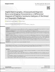
| dc.description.abstract | Objective: We aimed to investigate mammography (MG), ultrasound (US), and magnetic resonance imaging (MRI) findings of papillary breast carcinoma subtypes and to compare the diagnostic features and performance of the imaging method in distinguishing subtypes. Materials and Methods: Forty-two patients presenting with pathological diagnosis of 45 papillary carcinoma lesions, between 2014 and 2019, were included. Cases were assigned to five subgroups according to the latest World Health Organization (WHO) classification. The clinical characteristics (n = 45) and imaging features of each pathological subgroup were retrospectively related to imaging findings from US (n = 45), MG (n = 37), and breast MRI (n = 23), and further compared. Results: The finding of a palpable mass in all subgroups was more common than nipple discharge on clinical breast evaluation, and no significant difference was found between the subgroups. Irregular shape on MG (10/12, 83.3%, p = 0.039) and US (11/12, 91.7%, p = 0.039) was found more frequently in invasive micropapillary carcinoma (IMPC) compared to other subgroups. Circumscribed margins (4/5, 80%, p = 0.002) occurred more frequently in papillary ductal carcinoma in situ (pDCIS) and encapsulated papillary carcinoma (EPC) than in other subgroups (6/8, 75%, p = 0.002). Lower apparent diffusion coefficient (ADC) values were found in solid papillary cancer (SPC) than in other subgroups (ADC = 0.35 x 10(-3), p = 0.017). Conclusion: Radiological findings of papillary carcinomas overlap with each other. US and MRI are complementary when revealing specific morphological characteristics. | en_US |
| dc.language.iso | eng | en_US |
| dc.publisher | Galenos Yayincilik | en_US |
| dc.identifier.doi | 10.4274/ejbh.galenos.2022.2021-9-4 | |
| dc.rights | info:eu-repo/semantics/openAccess | en_US |
| dc.subject | Breast | en_US |
| dc.subject | Cancer | en_US |
| dc.subject | Magnetic Resonance Imaging | en_US |
| dc.subject | Mammography | en_US |
| dc.subject | Ultrasound | en_US |
| dc.subject | Invasive Micropapillary Carcinoma | en_US |
| dc.subject | Sonographic Findings | en_US |
| dc.subject | Ductal Carcinoma | en_US |
| dc.subject | Lesions | en_US |
| dc.subject | Features | en_US |
| dc.subject | Form | en_US |
| dc.title | Digital Mammography, Ultrasound and Magnetic Resonance Imaging Characteristics in Differential Diagnosis of Papillary Carcinoma Subtypes of the Breast and Diagnostic Challenges | en_US |
| dc.type | article | en_US |
| dc.relation.ispartof | European Journal of Breast Health | en_US |
| dc.department | Fakülteler, Tıp Fakültesi, Cerrahi Tıp Bilimleri Bölümü, Tıbbi Patoloji Ana Bilim Dalı | en_US |
| dc.identifier.volume | 18 | en_US |
| dc.identifier.issue | 2 | en_US |
| dc.identifier.startpage | 172 | en_US |
| dc.identifier.endpage | 181 | en_US |
| dc.institutionauthor | Öznur, Meltem | |
| dc.relation.publicationcategory | Makale - Uluslararası Hakemli Dergi - Kurum Öğretim Elemanı | en_US |
| dc.identifier.wos | WOS:000783230800009 | en_US |
| dc.identifier.trdizinid | 522381 | en_US |
| dc.identifier.pmid | 35445176 | en_US |